|
Breast Imaging ultrasound
If you touch a new thing, if your health provider touches something new, if you are over 40 years old or a direct family of you have suffrered breast cancer at any age, or you smoke, you can have a benefit from ultrasound imaging of your breast. No radiation, no unconfortable pressure. A complement of X rays procedures whenever the radiolopgical findings are inconclusive OR show something OR you feel something. DOES NOT substitute the X-ray procedure, it clarifies and complements it.
Our equipment
With a resolution of 14 MHz, we can diferentiate between two VERY SMALL adjacent points. Specially designed for fine diagnosis like breast imaging. To give you the idea: the transducers used for abdomen and pregnancy imaging range from 2 to 5 MHz, the usual breast transducer in most devices on the field are up to 12 MHz (commonly just 10 Mhz). The number shows in the pics included on your report like "L 8-10" or "L 7-12". Ours is L 14-6 (linear transducer ranging from 6 to 14 MHz harmonic imaging) REALLY HIGH RESOLUTION
|

|
|
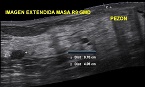
BREAST MASS, POSSIBLY BENIGN
|
|

TERMINAL DUCTAL LOBULAR UNIT
|
|

INTRAMMAMARY LIMPH NODE
|
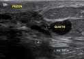
BREAST CYST
|
|
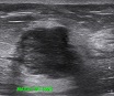
BIRADS IV
|
|
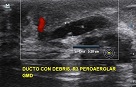
DEBRIS INSIDE DUCT
|
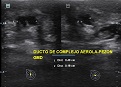
NIPPLE-AEROLAR COMPLEX
|
|
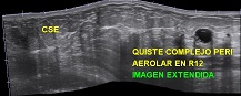
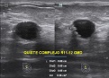
COMPLEX CYST
|
|
ZOOM IN
|
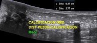
CALCIFICATION
|
|
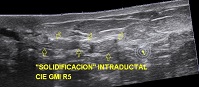
INTRADUCTAL MATTER
|
|
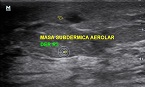
AEROLAR MASS
|
|
|
|

|
|
)
)
)
)
)
)
)
)
)
)
)
)