|
Imaging ultrasound
A prolongation of sight for human being that allows to see iside the body, the use of ultrasound is of paramount importance for the approaching on diverse health care situations for human kind.
Of course, the outstanding applications on Ob/Gyn are evident, however, almost no organ is an exception for investigation by means of imaging ultrasound. Faster, readily available and of course, cheaper than MRI or CT, and without the risks of radiation exposure or contrast media, the weakness of ultrasound usage is the fact of being highly operator-dependant. With our experience since 1985, of course that we have the knowledge (thanks God) to solve your questions, as not many of our colleagues presently have on the field
Our equipment
The following images talk by themselves. We count on devices for conventional ultrasound, color and doppler studies, duplex and triplex scan, endocavitary, 3D and "4D".
|

|
|

LIVER (COLOR DOPPLER)
|
|

3D GALLSTONES
|
|
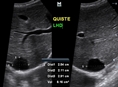
LIVER CYST
|

LIVER & GALLBLADDER
|
|
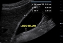
BILE DUCT STONE
|
|

DOPPLER. LIVER CYST
|

GALLBLADDER
|
|
GALLSTONES
|
|

GALLSTONES
|

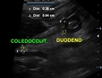
COMMON BILE DUCT
|
|
LIVER
|
|
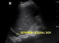
PLEURAL EFFUSION & LIVER
|
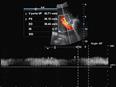
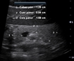
PORTAL VEIN BLOOD FLOW
|
|
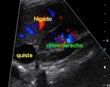
LIVER CYST & RIGHT KIDNEY DOPPLER
|
|
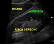
HEPATORRENAL RECESS
|
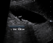
SMALL GALLSTONE (BILIARY SLUDGE)
|
|

BIG GALLSTONE &
|
|

DILATED COMMON BILE DUCT
|
PANCREAS
|
|
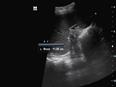
SPLEEN LENGHT
|
|

SPLEEN DOPPLER
|
|
|
|

|
|
)
)
)
)
)
)
)
)
)
)
)
)
)
)
)
)
)
)
)
)
)